2025
Using AI to spot Alzheimer's disease
Boston University highlighted our Nature Communications paper, AI-driven fusion of multimodal data for Alzheimer’s disease biomarker assessment.
Read moreResearch
Neurodegenerative diseases unfold over decades, rarely come alone, and look different in every person. The data we have about them is incomplete, scattered across clinics and cohorts, and hard to interpret, even for experts. We build AI for the hardest open problems this creates. We hold it to clinical standards: independent cohorts, autopsy-confirmed pathology, and side-by-side testing with clinicians. We make progress one well-validated step at a time.
The questions that matter most are the hardest to answer during life:
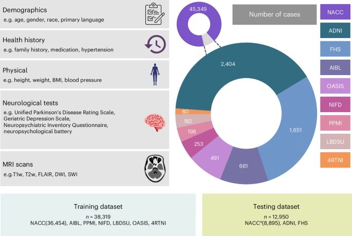
Today the answers need PET scans, spinal taps or autopsy, or they come years too late. Clues exist in MRI, cognitive tests, medical history, genetics and blood markers, but no patient has all of them, and every clinic and cohort collects a different subset on different equipment. Most AI assumes complete, uniform data, so it breaks down where it's needed most. The open problem is learning from data that is heterogeneous, partially observed and shifting from site to site, while holding the answers to the reference standard.
Code: Differential dementia diagnosis · Alzheimer's biomarker assessment · Multimodal AD dementia assessment · Interpretable AD classification · Disease-driven domain generalization · Dementia detection from voice · Speech features on GPU
Many diseases are ultimately confirmed under a microscope. Pathologists read tissue slides, electron micrographs show ultrastructure at nanometre scale, and fluorescence and other stains map specific molecules. For the neurodegenerative diseases we study, brain tissue examined at autopsy remains the reference standard. Digitizing these images has made them available to AI, but they are unlike everyday pictures. A single image can hold billions of pixels yet carry one label for the whole specimen, the informative structures may be tiny and scattered, and stains, microscopes and scanners vary from lab to lab. Expert reading is slow and scarce, and how tissue appearance relates to molecular state and clinical outcome is still largely unmapped.
Code: Graph-transformer for WSIs · Pathology + gene expression fusion · FourierMIL
Clinicians don't need another risk score at the end of a workup. They need help along the way: which test adds the most for its cost, who needs a specialist or a scan, and who will benefit from a new therapy or belongs in a trial. Every unnecessary test costs money and patient burden, and trials fail when they enroll the wrong people. To be actionable, AI has to reason about uncertainty and cost, and fit into a real clinical workflow.
General-purpose language models pass medical exams but stumble on the specifics of neurology and dementia care. They rarely cite evidence, and they can't read a brain MRI alongside a clinical note. Clinicians and researchers need models that know the field, reason over images and text together, and back every answer with sources that can be checked.
Code: PodGPT · Agentic medical QA
Press coverage, podcasts and highlights of our work.
Boston University highlighted our Nature Communications paper, AI-driven fusion of multimodal data for Alzheimer’s disease biomarker assessment.
Read moreAI-based differential diagnosis of dementia etiologies was included in the Nature portfolio on medical diagnostics.
Read moreThe Brink, BU's research news platform, covered our Nature Medicine model for differential diagnosis of dementia.
Read moreNature highlighted our Nature Medicine paper on an AI model for differential dementia diagnosis.
Read moreAn episode on building AI tools for dementia diagnosis, hosted by the Karen Toffler Charitable Trust.
Read moreA research briefing accompanying our Nature Medicine paper on AI for differential diagnosis of dementia.
Read moreA conversation on the possibilities of AI in neurological research and how these tools can improve efficiency in patient care.
Read moreOur Nature Communications (June 2022) model that uses multimodal data to assess Alzheimer's disease dementia was discussed on Clubhouse.
Read moreThe Brink covered our multimodal deep learning model for assessing Alzheimer's disease dementia.
Read moreA profile of our work with the Karen Toffler Charitable Trust.
Read moreThe Brink covered our interpretable deep learning model for Alzheimer's disease, published in Brain.
Read more